Epidural Steroid Injections

Author: Boqing Chen, MD, PhD Clinical Assistant Professor, Department of Physical Medicine and Rehabilitation, University of Medicine and Dentistry of New Jersey-New Jersey Medical School

Epidural steroid injections (ESIs) have been endorsed by the North American Spine Society and the Agency for Healthcare Research and Quality (formerly, the Agency for Health Care Policy and Research) of the Department of Health and Human Services as an integral part of nonsurgical management of radicular pain from lumbar spine disorders.

Radicular pain is frequently described as a sharp, lancinating, radiating pain, often shooting from the low back down into the lower limb(s) in a radicular distribution. Radicular pain is the result of a nerve root lesion and/or inflammation. Clinical manifestations of nerve root inflammation include some or all of the following: radicular pain, dermatomal hypesthesia, weakness of muscle groups innervated by the involved nerve root(s), diminished deep tendon reflexes, and positive straight or reverse leg–raising tests. In contrast to oral steroids, ESIs offer the advantage of a more localized medication delivery to the area of affected nerve roots, thereby decreasing the likelihood of potential systemic side effects. Studies have indicated that ESIs are most effective in the presence of acute nerve root inflammation.

The first documented epidural medication injection, which was performed using the caudal approach (see Approaches for Epidural Injections), was performed in 1901, when cocaine was injected to treat lumbago and sciatica (presumably pain referred from lumbar nerve roots).[1] According to reports, epidurals from the 1920s-1940s involved using high volumes of normal saline and local anesthetics. Injection of corticosteroids into the epidural space for the management of lumbar radicular pain was first recorded in 1952.

ESIs can provide diagnostic and therapeutic benefits. Diagnostically, ESIs may help to identify the epidural space as the potential pain generator, through pain relief after local anesthetic injection to the site of presumed anatomic pathology. In addition, if the patient receives several weeks or more of pain relief, then it may be reasonable to assume that an element of inflammation was involved in his or her pathophysiology. Since prolonged pain relief is presumed to result from a reduction in an inflammatory process, it is also reasonable to assume that during the period of this analgesia, the afflicted nerve roots were relatively protected from the deleterious effects of inflammation. Chronic inflammation can result in edema, wallerian degeneration, and fibrotic changes to the neural tissues.

In these authors’ opinion, ESIs are best performed in combination with a well-designed spinal rehabilitation program. In most cases, epidural injections should be considered as a treatment option after other treatment attempts (eg, physical therapy, including therapeutic exercise, manual therapy, and medications) have failed to improve the patient’s symptoms. However, ESIs may be indicated earlier in the treatment algorithm in some selected patients. Examples might include patients with medical contraindications to certain oral analgesics and patients whose pain severity substantially limits their ability to appropriately engage in therapeutic exercise.

A variety of approaches can be used to inject corticosteroids into the epidural space (see Approaches for Epidural Injections). For purposes of this article, the authors generally refer to all epidural steroid injections as ESIs, only specifying the specific type of approach if needed for a point of distinction or clarification.
Clinical Indications for Breast MRI

Amy Argus, MD; Mary C. Mahoney, MD

Mammography is the primary modality used for imaging the breast. However, it has known limitations. Dynamic contrast-enhanced breast magnetic resonance imaging (MRI) provides superior sensitivity to detect breast cancer and, when used in the appropriate clinical setting, it has become a useful adjunct to mammography.

Mammography is the primary modality used for imaging the breast. However, it has known limitations. Dynamic contrast-enhanced breast magnetic resonance imaging (MRI) provides superior sensitivity to detect breast cancer and, when used in the appropriate clinical setting, it has become a useful adjunct to mammography. Overlap in the MRI appearance of some benign and malignant diseases limits the specificity of breast MRI. The false-positive findings which result then prompt additional imaging and/or biopsies for benign disease. This, combined with the higher cost and limited availability of breast MRI vs. conventional imaging, requires appropriate use of the imaging modality. The American Cancer Society has outlined recommendations for the use of breast MRI for breast cancer screening and the American College of Radiology practice guidelines include 12 indications for the performance of breast MRI.[1,2]

[Read more…]

MRI Is the New Gold Standard for Excluding Cervical Spine Injury in Patients With Blunt Trauma

George D. Lundberg, MD

Cervical spine injury can have very serious consequences and can be difficult to diagnose. Five investigators in Wisconsin with a meta-analysis selected 5 prospective or retrospective published diagnostic protocol results that included magnetic resonance imaging. They studied reports of 464 victims of blunt trauma with clinically suspicious or unevaluatable cervical spines. Clinical follow-up was the gold standard. Sensitivity of MRI for cervical spine abnormalities was 97%, with a negative predictive value of 100% and zero false negatives; specificity was 99% with a positive predictive value of 94%; 97 patients, that is, 21%, had abnormalities by MRI that were not found by radiographs or CT. This study, reported in 2008 in The Journal of Trauma,[1] establishes MRI as the new gold standard for conclusively excluding cervical spine injury in a patient with blunt trauma.

Genetic Signature of Adult Gliomas and Correlation with MRI Features

Maria Grazia Bruzzone; Marica Eoli; Valeria Cuccarini; Marina Grisoli; Lorella Valletta; Gaetano Finocchiaro

In recent years the amount of information concerning the genetics and the biology of gliomas, and particularly of glioblastoma multiforme, increased steadily. Such an increase has been paralleled by the technological progress of MRI. The merging of these scientific areas, as summarized in this review, is helping the stratification of glioma patients for clinical trials and their clinical follow-up. Although available therapeutic options appear limited in number, it is likely that in the next 5 years, both as a consequence of the increased knowledge due to genomic sequencing of hundreds of glioblastoma specimens and to continuous improvements of MRI, new perspectives will be available for these patients, with a sizable impact on their prognosis.

Introduction

Gliomas, the most frequent tumors occurring in the CNS, are defined and graded on the basis of histological features, and pathology is fundamental to predict prognosis and guide the correct patient management.[1] However, pathological diagnosis can be rather subjective and allows considerable interobserver variability, especially in the case of gliomas with mixed histological features.[2] In addition, gliomas of identical histology may be associated with different genetic alterations. Therefore, owing to biological heterogeneity, the histological diagnosis and expected clinical outcome do not match in a significant number of patients and the histological examination does not distinguish tumors responding or not responding to the therapy. Throwing light upon individual biological alterations, molecular analyses may detect subsets of morphologically identical tumors with different clinical behavior (diagnostic markers), describing their prognosis more effectively (prognostic markers).[3] Moreover, molecular biological studies may lead to the discovery of gene-based predictors of therapeutic response, helping to guide more rationally currently available therapies (predictive markers).[4] At present few tumor biomarkers are available for gliomas and it is sometimes unclear how to incorporate molecular genetic information into clinical practice. Differences in study design, patient and specimen characteristics, assay methods and statistical analysis make different studies poorly comparable and also make it difficult to understand the context in which the conclusion should be applied.[5]

[Read more…]

MRI Guides Diagnostic Approach for Ischaemic Stroke

M A Kumar; H Vangala; D C Tong; D M Campbell; A Balgude; I Eyngorn; A S Beraud; J M Olivot; A W Hsia; R A Bernstein; C A Wijman; M G Lansberg; M Mlynash; S Hamilton; M E Moseley; G W Albers

Background and aim Identification of ischaemic stroke subtype currently relies on clinical evaluation supported by various diagnostic studies. The authors sought to determine whether specific diffusion-weighted MRI (DWI) patterns could reliably guide the subsequent work-up for patients presenting with acute ischaemic stroke symptoms.
Methods 273 consecutive patients with acute ischaemic stroke symptoms were enrolled in this prospective, observational, single-centre NIH-sponsored study. Electrocardiogram, non-contrast head CT, brain MRI, head and neck magnetic resonance angiography (MRA) and transoesophageal echocardiography were performed in this prespecified order. Stroke neurologists determined TOAST (Trial of Org 10172 in Acute Stroke Treatment) classification on admission and on discharge. Initial TOAST stroke subtypes were compared with the final TOAST subtype. If the final subtype differed from the initial assessment, the diagnostic test deemed the principal determinant of change was recorded. These principal determinants of change were compared between a CT-based and an MRI-based classification schema.
Results Among patients with a thromboembolic DWI pattern, transoesophageal echocardiography was the principal determinant of diagnostic change in 8.8% versus 0% for the small vessel group and 1.7% for the other group (p<0.01). Among patients with the combination of a thromboembolic pattern on MRI and a negative cervical MRA, transoesophageal echocardiography led to a change in diagnosis in 12.1%. There was no significant difference between groups using a CT-based scheme.
Conclusions DWI patterns appear to predict stroke aetiologies better than conventional methods. The study data suggest an MRI-based diagnostic algorithm that can potentially obviate the need for echocardiography in one-third of stroke patients and may limit the number of secondary extracranial vascular imaging studies to approximately 10%.

[Read more…]

The Clinical Use of Structural MRI in Alzheimer Disease

Giovanni B. Frisoni; Nick C. Fox; Clifford R. Jack; Philip Scheltens; Paul M. Thompson

Structural imaging based on magnetic resonance is an integral part of the clinical assessment of patients with suspected Alzheimer dementia. Prospective data on the natural history of change in structural markers from preclinical to overt stages of Alzheimer disease are radically changing how the disease is conceptualized, and will influence its future diagnosis and treatment. Atrophy of medial temporal structures is now considered to be a valid diagnostic marker at the mild cognitive impairment stage. Structural imaging is also included in diagnostic criteria for the most prevalent non-Alzheimer dementias, reflecting its value in differential diagnosis. In addition, rates of whole-brain and hippocampal atrophy are sensitive markers of neurodegeneration, and are increasingly used as outcome measures in trials of potentially disease-modifying therapies. Large multicenter studies are currently investigating the value of other imaging and nonimaging markers as adjuncts to clinical assessment in diagnosis and monitoring of progression. The utility of structural imaging and other markers will be increased by standardization of acquisition and analysis methods, and by development of robust algorithms for automated assessment.

Introduction

Clinical and neuropathological studies have greatly advanced our knowledge of the pathophysiology and progression of Alzheimer disease (AD). This disease is associated with progressive accumulation of abnormal proteins (amyloid-β [Aβ] and hyperphosphorylated tau) in the brain, which leads to progressive synaptic, neuronal and axonal damage. Neurobiological changes occur years before symptoms appear, with a stereotypical pattern of early medial temporal lobe (entorhinal cortex and hippocampus) involvement, followed by progressive neocortical damage.[1,2] The delay in emergence of the cognitive correlates of these changes suggests that the toxic effects of tau and/or Aβ progressively erode ‘brain reserve’ until a clinical threshold is surpassed and amnestic symptoms develop. For example, amnestic mild cognitive impairment (MCI)—memory disturbance in the absence of dementia—is followed by more-widespread cognitive deficits in multiple domains until a disability threshold is reached and traditional diagnostic criteria for probable AD are fulfilled.[3] The prospect of disease-modifying drugs that can slow or prevent disease progression has prompted increased interest in identifying individuals with AD earlier and more accurately.

[Read more…]

What is MRI?

Magnetic Resonance Imaging uses friendly magnetic fields to produce an unparalleled view inside the human body. MRI has become the diagnostic imaging modality of choice for most malignancies and neurological diseases of the brain and spine including:

  • Brain tumors
  • Alzheimer’s Disease
  • Cervical cancer

The technology’s ability to generate, non-invasively, superb anatomical details of both bone and soft tissues has made MRI the preferred modality for most orthopedic applications including imaging of:

  • Knee, hip, shoulder, wrist, elbow
  • Ligaments and tendons
  • Back pain and failed back syndrome

Do I need a Prescription for an MRI?

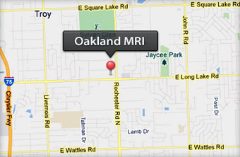
You will need a prescription for an MRI. If you have reason to believe that an MRI would be beneficial in diagnosing your physical condition more accurately, discuss it with your doctor. Feel free to have your physician call Oakland MRI to talk about your case. If your physician agrees that an MRI exam would be beneficial, he or she can refer you to Oakland MRI for a scan. You may download our Prescription Form for your convenience.

How do I prepare for my MRI exam?

Good news – no special preparation is needed. Eat and take any prescribed medication as usual, unless your doctor tells you otherwise.

One important thing to remember, though: MRI and metal do not mix.

An MRI system has a powerful magnet inside, which is why you need to follow these guidelines:

Tell your physician and the MRI staff if you have a pacemaker, prosthesis, surgical clips, metal implants, or any other metal objects in your body. Some implants, such as a pacemaker, may be affected by an MRI exam.
Leave metal or magnetized objects at home or give them to the MRI staff for safekeeping when you arrive for the exam. Items that might be affected by the magnet include watches, coins, keys, bobby pins or other hair clips, pocketknives, and credit cards.

Avoid wearing eye makeup because metal flakes or slivers are found in some eye shadows.
Let the technologist know if you work around metal finishing or grinding equipment. It’s important to keep the eye area free of any metallic particles.

Finally, if you have additional questions or want more information before your exam, please don’t hesitate to contact your physician or the staff of the MRI facility.

What can I expect during my MRI exam?

The MRI exam itself is painless – you won’t feel a thing. You may notice a slight knocking noise as each image is taken, however, so let your technologist know if you are particularly sensitive to sound.

As with any exam, the hardest part is to be patient. Just relax and remain still. The MRI staff will keep you informed every step of the way. Remember, they are there to assist you. Should you become uncomfortable, need help, or have questions at any point during your exam, just say so. There is a built-in intercom in the system so you can talk with the staff.

The length of your exam depends on the type of study your doctor has ordered. In many cases, MRI patients are done and on their way home within an hour.

After your exam, the technologist will take you back to the preparation room to collect your things. That’s all there is to it.