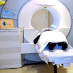
 People with pacemakers or implantable defibrillators have long been told they can’t undergo MRI scans. But a new study suggests that it can be safely done — under the right conditions.
The study, published in the Feb. 23 issue of the New England Journal of Medicine, focused on patients with standard heart devices not designed to be MRI-compatible.
The study found that even for them, an MRI can be safely performed, when a specific protocol is followed.
“I think this really opens a door for these patients to have an MRI when medically indicated,” said lead researcher Dr. Robert Russo, of the Scripps Research Institute, in La Jolla, Calif.
The big caveat, though, is that patients in the study were all screened and went through a specific protocol.
An expert in cardiac devices — a doctor, physician’s assistant or nurse practitioner — had to be present during the MRI. Each patient’s device was reprogrammed before the MRI scan, then restored to the original settings afterward. And during that time, patients’ blood pressure, blood oxygen and heart rhythm were all monitored.
The bottom line is, patients cannot safely get an MRI at just any center, according to Dr. Emanuel Kanal, director of magnetic resonance services at the University of Pittsburgh Medical Center.
“Yes, some patients can be scanned, under specific circumstances,” said Kanal, who was not involved in the study.
But he stressed that it should only be done at centers with the necessary expertise in managing patients with these heart devices.
MRI scans are done for a wide range of reasons, to help diagnose anything from tumors to bone and joint injuries, to internal bleeding. Unlike CT scans, an MRI scan doesn’t involve radiation, Russo pointed out — and it can sometimes give information that other types of imaging cannot.
So, he said, it’s important that people with heart devices be able to undergo an MRI scan, if possible.
But because an MRI scan produces a powerful magnetic field, there has long been concern about its safety for heart-device patients. One of the biggest worries, Russo said, is that it could overheat the device wires, possibly injuring the heart muscle or altering how the device paces the heart.
So for years, people with pacemakers and implantable cardioverter defibrillators (ICDs) have been warned against undergoing an MRI.
In more recent years, manufacturers have developed so-called “MRI-conditional” heart devices, which are designed to minimize the potential risks of the scans.
Still, Russo said, many people have conventional devices — an estimated 2 million in the United States alone. So his team looked at whether such patients can safely have an MRI, with the right precautions in place.
The study involved more than 1,200 patients with standard pacemakers or ICDs. They underwent a total of 1,500 MRI scans at 19 centers across the United States.
All of the scans were done using the same protocol. Each patient’s device was “interrogated” — tested non-invasively — before the MRI, then reprogrammed accordingly. If possible, it was set to a “no-pacing” mode. But if patients had symptoms in that mode, their device was programmed a different way.
After the MRI, the device was restored to its original settings, then tested again to make sure it was working properly.
None of the patients, the study found, had a “failure” in their device or its wires during the MRI. And none suffered a dangerous heart-rhythm disturbance.
Six patients did have atrial fibrillation or atrial flutter — a quivering in the heart’s upper chambers. But each case was short-lived and stopped on its own, the study authors said.
One ICD patient needed to have the device generator immediately replaced after the MRI.
That, Russo said, was because “someone left the shock function on” before the scan. The device went into non-functional mode during the MRI, then couldn’t be tested afterward.
But overall, Russo said, “we didn’t find any true risk that should stop these patients from having a medically indicated MRI.”
None of the patients had an MRI of the chest, however — so it’s not clear whether the findings apply to those scans, the researchers acknowledged.
To Kanal, the take-away for patients is this: “They’re not necessarily precluded from having any MRI study of their body — even if they have a device that’s not MRI-conditional.”
But he underscored the importance of having the scan done at the right center. He also pointed to one way patients can do their homework: Find out whether your MRI center has personnel certified by the American Board of Magnetic Resonance Safety.
Dr. David Wilber is editor-in-chief of the journal JACC: Clinical Electrophysiology. He called the study findings “good news,” adding that “this protocol has been incorporated at many medical centers, and these findings confirm that it is quite safe.”
However, given the personnel required, “it will probably be limited to relatively large centers,” and may or may not be covered by patients’ insurance whose devices aren’t “MRI-conditional.”
The study was partly funded by St. Jude Medical, Biotronik and Boston Scientific, all makers of heart devices.
SOURCES: Robert Russo, M.D., adjunct assistant professor, molecular and experimental medicine, Scripps Research Institute, La Jolla, Calif.; Emanuel Kanal, M.D., director, magnetic resonance services, and professor, radiology and neuroradiology, University of Pittsburgh Medical Center, Pittsburgh; David Wilber, M.D., editor-in-chief, JACC: Clinical Electrophysiology; Feb. 23, 2017 New England Journal of Medicine

