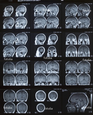
 Three-fourths of patients with Alzheimer’s disease or frontal-lobe degeneration had MRI-detected biomarker levels that correlated with the diagnoses, suggesting MRI has potential as a screening tool for the conditions, investigators reported.
MRI-predicted values for total tau and β-amyloid ratio (tt/Aβ) in gray matter correctly pinpointed the diagnosis in 75% of patients with genetically or neuropathologically confirmed diagnoses, according to Corey McMillan, PhD, of the University of Pennsylvania in Philadelphia, and colleagues. Predicted values also had good correlation with actual tt/Aβ measured in cerebrospinal fluid (CSF), they said.
The findings are consistent with previous in vivo and autopsy studies of CSF tt/Aβ, the group reported in the Jan. 8 issue of Neurology.
“Specifically, MRI-predicted and actual CSF tt/Aβ values are highly correlated, predicted tt/Aβ accurately defines the anatomic distribution in Alzheimer’s disease and frontotemporal lobar degeneration (FTLD), and predicted tt/Aβ values are reasonably accurate for classifying individual patients as having Alzheimer’s disease or FTLD pathology,” they wrote.
“This study establishes empirical evidence that an MRI-based technique can predict a single, biologically valid level of CSF tt/Aβ. This may contribute to diagnosis and treatment trials of neurodegenerative conditions by screening for individuals requiring a more invasive diagnostic lumbar puncture,” they added.
Measurement of tt/Aβ in CSF has demonstrated diagnostic accuracy for distinguishing between Alzheimer’s disease and FTLD. However, the measurement requires lumbar puncture, which is invasive and problematic in situations that require repeated measurements, such as a clinical trial. An accurate, non-invasive alternative is needed, the authors noted.
Volumetric MRI has shown potential as an alternative to assessment of CSF tt/Aβ because of its ability to capture neuroanatomical features that distinguish Alzheimer’s disease from FTLD.
The authors performed a prospective study to compare MRI-predicted versus actual CSF tt/Aβ in 185 patients with clinically diagnosed neurodegenerative disease. All patients underwent lumbar puncture, and CSF tt/Aβ showed a profile consistent with Alzheimer disease in 88 cases and other conditions in the remaining 97 patients.
The patients underwent volumetric MRI an average of 5 months after lumbar puncture.
The authors estimated MRI-predicted CSF tt/Aβ on the basis of the extent of gray matter atrophy. Comparison of predicted and actual CSF tt/Aβ showed significant correlation between the two measures (P<0.0001).
Next, investigators examined neuroanatomic features of the patients’ gray matter to develop profiles associated with measured CSF tt/Aβ. Lower actual values for CSF tt/Aβ, indicative of FTLD, were associated with reduced gray matter density in the ventromedial prefrontal cortex, orbital frontal cortex, insula, thalamus, and anterior temporal cortex.
Higher CSF tt/Aβ values, associated with Alzheimer’s disease, were associated with reduced gray matter density in posterior regions, including superior parietal cortex, precuneus, and occipital association cortex.
“A regression revealed a very similar distribution of reduced gray matter density associated with tt/Aβ levels predicted by MRI,” the authors wrote. “Lower predicted tt/Aβ values, consistent with FTLD, were related to reduced gray matter density in frontal regions … by comparison, higher predicted tt/Aβ values, consistent with Alzheimer’s disease, were related to reduced density in posterior gray matter regions.”
The investigators compared the predicted and actual tt/Aβ in a subset of 32 patients with known pathology (21 with FTLD, 11 with Alzheimer’s), as determined by genetic mutations or detailed neuropathologic studies. Using a cutoff of -1.38 for actual tt/Aβ resulted in 91% sensitivity and 81% specificity for Alzheimer’s disease and overall classification accuracy of 84%.
The authors reported that 17 patients were correctly identified as having FTLD, and 10 patients with Alzheimer’s disease were classified correctly on the basis of actual tt/Aβ.
Using the same cutoff value, investigators repeated the calculations for MRI and found 75% overall accuracy for classification of the patients: 17 of 21 patients with FTLD and seven of 11 with Alzheimer’s disease.
McMillan’s group cautioned that the trajectory of disease may be a limiting factor for MRI-based CSF screening.
“While we demonstrate that the distribution of gray matter atrophy in Alzheimer’s disease and FTLD is highly related to a distinct range of CSF tt/Aβ values, these biological changes may occur at different stages in the disease course,” they said.
The authors of an accompanying editorial praised the work as another step forward in defining MRI’s role in pathologic diagnosis.
“The results of McMillan et al are impressive,” wrote Christian Habeck, PhD, of Columbia University in New York City and Jennifer Whitwell, PhD, of the Mayo Clinic in Rochester, Minn. “The clinical diagnoses of the patients with Alzheimer’s disease and FTLD overlapped substantially, demonstrating utility for predicting pathology in clinically atypical patients in which diagnosis is the most challenging.”
By Charles Bankhead, Staff Writer, MedPage Today

